




Quantitative analysis of leukocyte phagcytosis in 100 µl of whole blood under optimal conditions without any disturbance by isolation procedures
A. Sample Preparationfor opsonized E.coli-FITC.Simple as you know it from immunofluorescence analysis: 1.Labeling |
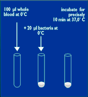 |
2. Quenching |
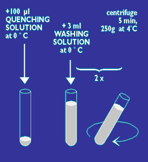 |
3. Lysing and Fixation |
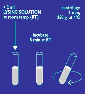 |
4. Washing and DNA Staining |
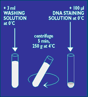 |
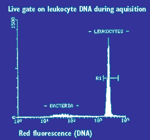
B. MeasurementGating on leukocytes after DNA staining:Discrimination of bacteria, single leukocytes and doublets |
 |
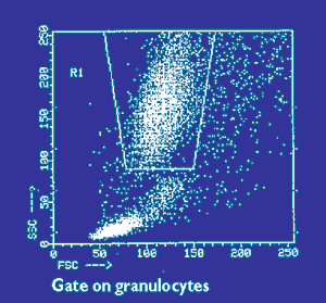
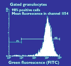
C. Data EvaluationGate on monocytes and granulocytes and determine the percentage of phagocytosing cells and their phagocytic activity (= mean fluorescence = number of bacteria per cell) |
|
Report your results |
![[Back]](../../bilder/ba_b.gif)
![[Contact]](../../bilder/co_b.gif)
![[Description]](../../bilder/de_b.gif)
![[Handling]](../../bilder/ha_b.gif)